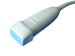
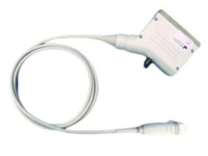
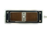
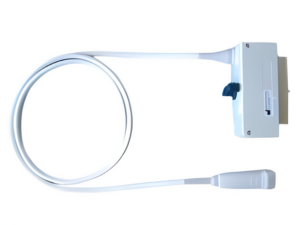
Phased Array Probe | S3-21311A Compatible | 1-3MHz
The S3-21311A model is a phased array probe. This transducer comes with a smaller footprint and low frequency which allows medical practitioners to perform scans of the smaller spaces. It produces the scan results with a narrow beam at the top that widens at the end which covers a wider…
| Compatible Systems |
Philipps HP Sonos 7500 series etc |
|---|---|
| Product Reference |
UPDP S3-21311A |
| Shipping Method |
DHL, FedEx, etc |
| Payment Method |
Paypal, T/T |

- Mechanical focus of ultrasound beam
- Model-specific materials and dimensioning
- Electrical insulation
- Fluid barrier
- Single or multilayer materials, must be ISO 10993 compliant
- Maximizes transmission of energy from acoustic array to tissue by reducing reflection and increases spectrum of frequencies(bandwidth) emitted by transducer
- Consists of one to seven different layers based on transducer model
- Generally ¼ wavelength of the array’s center frequency in thickness
- Converts electrical energy to mechanical energy (pressure wave) and vice versa
- Consists of several to thousands of individual elements
- Reduces the effects of electromagnetic interference
- Typically surrounds the array, the backing material and scan head electronics
- Dampens crystal vibrations to reduce pulse duration, which increases axial resolution
- Flexible circuit board that connects the inter connect board to the individual elements
- Bridge between individual coaxial cables and flex circuit
- May include multiplexing circuitry, as well as beam forming circuitry
- Bridge between the connector electronics and the scan head electronics
- Internal signal wire enclosed within a braided shield
- Typically the diameter of a hair
- Model-specific length, impedance and capacitance
- Reduces stress on main and coaxial cables
- Protects main transducer cable
- Contains several to hundreds of individual coaxial wires
- Model specific shielding to minimize the effects of electromagnetic interference
Ultrasound imaging remains a cornerstone in medical diagnostics, cherished for its non-invasive nature and versatility. The heart of this technology is the ultrasound probe, a critical device that converts electrical energy into sound waves and vice versa, revealing detailed images of the body’s internal structures. From linear to endorectal, micro convex and phased array, from low frequency to high frequency, Ultrasound Probe Direct stays committed to meet every clinical need and preference, enhance your diagnosis accuracy and improve patient’s care and experience.
Key MaterialsThe choice of materials for ultrasound probes has seen significant refinement, driven by the need to meet highest performance criteria:
Piezoelectric Properties: This remains the core characteristic, enabling the conversion between electrical and mechanical energy. Lead zirconate titanate (PZT) continues to be widely used due to its excellent piezoelectric capabilities, cost-effectiveness, and manufacturability.
Alternative Piezoelectric Materials: In addition to PZT, materials such as polyvinylidene fluoride (PVDF) and barium titanate are gaining popularity. PVDF, a polymer, offers benefits in terms of weight and flexibility but comes with a higher cost. Barium titanate has superior piezoelectric properties but poses challenges in terms of cost and manufacturing complexity.
Beyond the piezoelectric core, several components are crucial to the functionality of an ultrasound transducer:
Backing Material: This component, positioned behind the piezoelectric material, dampens vibrations and enhances sound wave transmission efficiency.
Matching Layer: Located in front of the piezoelectric material, it minimizes sound wave reflection by matching the transducer's acoustic impedance to that of the body.
Housing and Cabling: These elements protect the transducer, facilitate its attachment to the imaging probe, and ensure efficient electrical signal transmission between the transducer and the imaging system.
The construction of an ultrasound probe is a complex process that includes:
Transducer Assembly: This involves bonding the piezoelectric material to the backing, applying the matching layer, and mounting it within the protective housing.
Electronic Integration: It incorporates signal processing units, power supply components, and control circuitry.
Quality Testing: Rigorous testing is conducted to ensure the transducer, electronics, and overall imaging system function effectively.
Sterilization: Probes undergo sterilization to meet healthcare standards, a critical step for medical usage.
- Our ultrasound probes are shipped from our strategically located global distribution centers to ensure timely delivery to our customers round the world.
- The destination, shipping mode, and order weight are among the variables that go into calculating shipping costs. You can get an accurate shipping quote during the website's checkout process.
- Our customer support team is always here to assist you with your order, whether you have questions about the product, shipping, or payment. They can be contacted by phone, email, or live chat on our website.
- In order to guarantee product quality and performance, our ultrasound probes come with a standard warranty period. The product documentation contains specifics about the warranty duration, which may differ based on the product. Please get in touch with our support staff if you need more details or have questions about warranty coverage.
- Using the search bar on the home page of our website, you can quickly look for a specific ultrasound probe. To display relevant results, just enter the product name or model number.
- Please get in touch with our customer service team if you can't find a specific product on our website. We are always glad to assist you in locating the product or provide alternative solutions.
- Using the ADD TO QUOTE button on the product detail page of our website, you can quickly get a quote for your ultrasound probe products or services at any time. To submit your request, just click the SEND button on directed Request A Quote page with all selected products included in a filled form.
- Our ultrasound probes are available for order by Healthcare providers, hospitals, veterinary clinics, research facilities, and licensed distributors.
- It's simple to sign up on our website in advance or during the checkout process. Order tracking, purchase history, and expedited checkout are just a few advantages of creating an account, even though it's not required.
- Yes, you can get in touch with our committed sales team over the phone to place an order. They will help you with the ordering process and respond to any queries or worries you might have.
- Periodically, we run sales and promotions for our ultrasound probes. To receive updates on new deals and discounts, make sure to sign up for our newsletters or visit our website frequently.
- You will receive an email with a tracking number as soon as your order ships. Using this tracking number, you can check the location and status of your package on the carrier's website.
- Orders can usually be canceled before they are processed for shipping, provided that they happen within a specific window of time after the order is placed. If you would like to cancel your order, please get in touch with our customer support team right away.
- Our customer support team is always here to assist you with your order, whether you have questions about the product, shipping, or payment. They can be contacted by phone, email, or live chat on our website.
- In order to guarantee product quality and performance, our ultrasound probes come with a standard warranty period. The product documentation contains specifics about the warranty duration, which may differ based on the product. Please get in touch with our support staff if you need more details or have questions about warranty coverage.
Legal Notice: This medical device is a healthcare regulated product. In compliance with the regulation, this products bears the CE marking. Denomination: Compatible Transducer. Purpose: Ultrasound Medical Imaging System. Instructions: Read carefully the user manual. Consult a Healthcare Professional. Class: 2a.
What is an ultrasound?
An ultrasound is an imaging test that uses sound waves to make pictures of organs, tissues, and other structures inside your body. It allows your health care provider to see into your body without surgery. Ultrasound is also called ultrasonography or sonography. Ultrasound images may be called sonograms.
Ultrasound can be used to treat certain medical conditions. But it’s mostly used to help:
- Monitor the health and development of an unborn baby during pregnancy. Pregnancy ultrasound can help check if your baby is growing normally. It can screen for certain conditions, such as birth defects that can be seen in images. It can also check for pregnancy problems. For example, an ultrasound can show if your placenta (the organ that brings oxygen and nutrients to the baby) is in the right position. Pregnancy ultrasound may also be called “prenatal ultrasound,” “fetal ultrasound,” or “obstetrical ultrasound.”
- Diagnose the cause of a wide variety of medical conditions. Ultrasound is best used to learn about conditions that involve soft tissues, such as organs, glands, and blood vessels. Diagnostic ultrasound may be used if you have signs or symptoms of a problem, and an ultrasound may help diagnose or rule out possible causes.
- Guide certain biopsy procedures. Some biopsies use a needle to remove a sample of fluid or tissue from the body for testing. An ultrasound can find the abnormal area and guide the needle to the right place to collect the sample.
There are different types of ultrasounds. One type, called Doppler ultrasound, can show movement in your body. For example, it can show your heart beating and the speed and direction of blood flowing through your blood vessels. It can also show the beating heart and movement of an unborn baby. Another type of ultrasound can create 3-D (three-dimensional) images.
Other names: sonogram, ultrasonography, pregnancy sonography, fetal ultrasound, obstetric ultrasound, diagnostic medical sonography, diagnostic medical ultrasound
What is it used for?
A pregnancy ultrasound may be used to:
- Check the size, position, heart rate, and age of the unborn baby
- See if there is more than one baby
- Screen for:
- Genetic disorders, such as Down syndrome
- Birth defects in the heart, brain and spinal cord, or other parts of the body
- Check the amount of amniotic fluid (the liquid in the sac surrounding an unborn baby) and the location of the placenta
- Guide the collection of test samples taking during amniocentesis and chorionic villus sampling (CVS)
Diagnostic ultrasound has many uses. For example, ultrasound can help:
- Find the cause of pain, swelling, and other symptoms
- Look for blockages, growths, and structural problems in organs, glands, and blood vessels
- Tell the difference between cysts (fluid-filled sacs) and solid tumors
Ultrasound may help diagnose medical conditions that involve many parts of the body, such as the:
- Heart and heart valves
- Blood vessels
- Organs in the abdomen (belly), including the liver, gallbladder, pancreas, or spleen
- Organs in the pelvis, including the urinary tract, and male and female reproductive organs
- Thyroid and parathyroid glands
- Kidneys
- Breasts
- Brain, spine, and hips in infants
Why do I need an ultrasound?
If you’re pregnant, it’s common to have a routine ultrasound between weeks 18 and 22 of pregnancy to check on the health of your baby. If your provider suspects a problem, you may need an ultrasound at other times during your pregnancy.
You may need a diagnostic ultrasound if you have signs or symptoms of certain types of medical conditions and your provider needs to look inside your body to help find the cause. If you’re having a needle biopsy to remove fluid or tissue for a test, you may have an ultrasound as part of the procedure.
What happens during an ultrasound?
An ultrasound is often done by a sonographer, a health care professional who has special training to do ultrasound exams. Ultrasounds may be done in different ways depending on the part of your body that’s being examined. Most ultrasound exams include these general steps:
- You will remove clothing from the area that will be examined and lie on a table.
- The sonographer will spread a special gel on your skin over the area that will be examined.
- The sonographer will hold a wand-like device, called a transducer, and move it across your skin. The device sends sound waves into your body. The gel prevents air from getting between the device and your skin, which would block the soundwaves from entering your body. The sound waves are a very high pitch, so you can’t hear or feel them.
- The soundwaves “bounce” off the structures inside your body. The ultrasound device picks up the echoes and turns them into images on a computer screen. You may be able to see the images on the screen during your exam.
- After the exam is over, the sonographer will wipe the gel off your skin.
For certain ultrasound exams, the ultrasound device is placed inside a body opening to get a clearer image. Depending on the organs being checked, the device may be placed in the:
- Vagina. This is called a transvaginal ultrasound. It helps view the uterus and ovaries.
- Rectum. This is called a transrectal ultrasound. It’s usually done to view the prostate gland.
- Esophagus (the tube that connects your mouth and stomach). This is called a transesophageal echocardiogram (TEE). It’s done to get clear images of the heart.
Will I need to do anything to prepare for the test?
Preparations for an ultrasound exam depend on what part of your body is being checked. Some ultrasound exams require no preparation at all. Your provider will tell you if you need to prepare for your ultrasound and what to do.
For example, if you’re having an ultrasound to view your urinary tract, you may need to drink water so that you have a full bladder. You will have to hold your urine (pee) until the test is over. For other ultrasounds, you may need to fast (not eat or drink) for several hours before your test.
Are there any risks to the test?
Ultrasound has not been linked to any health harms. It’s generally considered safe when trained sonographers use it correctly. Ultrasound doesn’t use ionizing radiation like x-rays use, which makes it safer than x-ray. That’s why ultrasound is the most widely used medical imaging method for viewing an unborn baby during pregnancy.
But in certain cases, ultrasound can affect fluids and tissues in the body. That’s why most medical experts recommend using ultrasound only when it’s needed to provide important medical information.
What do the results mean?
The results of your ultrasound depend on the type of ultrasound you had. Your provider can explain what your results mean for your health.
If you had a pregnancy ultrasound, normal results mean that your baby appears to be developing normally. But an ultrasound can’t guarantee you’ll have a healthy baby. If your results weren’t normal, you may need more tests, including another ultrasound exam.
Talk to [representative] to see if [product] is right for you.
Phone:
Email:
Online Chat: 24 hours available






Reviews
There are no reviews yet.