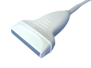
How to Choose the Right Ultrasound Probe: The Definitive Guide
Discover how to select the best ultrasound probes for your medical practice. The guide ensures you choose the right probe for optimal diagnostic accuracy and patient care.
Introduction
Any medical practice must be careful to consider which the ultrasound probe should be used. Because it affects diagnostic accuracy and patient care directly. The right medical probe can make sure the clear and exact ultrasound imaging, which is necessary for precise diagnosis and efficient treatment planning. Certain probe properties are important for certain medical applications, such as footprint and frequency. The reason is that these probe properties can help doctors to get the best images of the targeted body areas.
Through using the appropriate ultrasound probe, doctors will get the quality of medical evaluations and reduce diagnostic errors and unnecessary treatments. Therefore, the right probe can improve patient outcomes, increase overall patient safety and satisfaction. What’s more, a suitable medical probe provides faster and more accurate tests, which enhances the efficiency of medical practices. In a word, purchasing the right ultrasound probes is an investment in the quality of treatment your clinic provides.
Understanding Ultrasound Probes
Medical ultrasonography machines require ultrasound probes, or transducers. These handheld devices send high-frequency sound waves into the body and bounce against organs, tissues, and other structures. The probe converts these reflected sound waves into electrical signals that the ultrasound machine uses to create real-time bodily images.
Ultrasound probes are essential for medical imaging diagnosis and treatment. Healthcare workers utilize them to visualize organs, blood arteries, and tissues to diagnose problems, monitor conditions, and guide surgeries. For superficial imaging, linear probes are used, convex probes for abdominal exams, and phased array probes for cardiac exams.
Since they don’t employ ionizing radiation, ultrasound probes are safe, non-invasive, and fast. Their versatility and efficacy make them essential in obstetrics, gynecology, cardiology, radiography, and emergency medicine. Clinicians can improve patient care and diagnoses by using the right probe.
Different Types of Ultrasound Probes
Ultrasound probes, also known as transducers, come in various types, each designed for specific medical applications. Here are some of the most common types:
- Linear Probes:
A linear probe is characterized by its rectangular shape and its ability to generate sound waves at a high frequency. Because they have such a high resolution, they are perfect for imaging structures that are only surface. Vascular investigations, musculoskeletal imaging, thyroid screenings, and imaging of tiny parts (such as breast and testicular exams) are examples of common applications. In most cases, the frequency range is between 7.5 to 17 MHz

- Convex (Curved Array) Probes:
The convex probes have a curved surface, which enables them to have a broader field of view and to detect lower frequency sound waves. The fields of obstetrics, gynecology, and abdominal imaging are prominent applications for these devices. Because they have a wider field of view, they are excellent for structures that are deeper. In most cases, the frequency range is between 2 to 7 MHz.

- Phased Array Probes:
Phased array probes are small and have a piezoelectric element arranged in a compact grid. They can steer the sound beam electronically, allowing for dynamic focusing and a small footprint. These probes are primarily used in cardiac imaging (echocardiography) due to their ability to capture images between the ribs. They are also used in emergency medicine and for transcranial Doppler studies. Typically, between 1 to 5 MHz.

- Endocavitary Probes:
Designed for use in internal investigations, endocavitary probes are characterized by their long and thin shape. The purpose of inserting them into body cavities is to provide a more detailed image of the internal architecture. In the fields of obstetrics and gynecology, transvaginal scans are frequently performed, while in the field of urology, transrectal prostate exams are performed. In most cases, the frequency range is between 5 to 9 MHz.
- 3D/4D Probes:
These probes have the ability to capture photos in three dimensions as well as images in four dimensions in real time. Arrays can be either linear or curved, depending on need. Both in the field of cardiology and in the field of obstetrics, where it is utilized for thorough fetal imaging, as well as for volumetric heart examinations. Variable frequency range is determined by the type of base probe, which can be either linear or convex.
- Intraoperative Probes:
Probes that are meant to be used during surgical procedures are called intraoperative probes. They can be used directly on organs and tissues, and they are frequently capable of being sterilized. Implemented in a variety of surgical procedures, such as those involving the liver, kidneys, and vascular system, to give real-time imaging and to direct surgical decision-making. In most cases, the frequency range is between 5 to 10 MHz.
Factors to Consider When Selecting an Ultrasound Probe
When selecting the appropriate ultrasonic probe, it is crucial to consider several key factors to ensure it meets the requirements of your medical practice. Here are the essential aspects to evaluate:
Frequency
Frequency is measured in megahertz (MHz), which represents the number of sound wave cycles per second. Lower frequencies provide deeper penetration but with lower resolution, while higher frequencies offer better resolution but with less penetration.
- Low-Frequency Probes (2 to 5 MHz): Ideal for imaging deeper regions such as the liver, heart, and abdomen.
- High-Frequency Probes (7.5 to 17 MHz): Best suited for imaging superficial structures like the breast, thyroid, and musculoskeletal system.
Footprint
The size and shape of the probe’s head affect its field of view and contact area.
- Compact Footprint Probes: Ideal for confined or hard-to-reach areas, such as imaging the heart between the ribs.
- Large Footprint Probes: Perfect for imaging large areas like the abdomen.
Application
The specific medical use for which the probe is intended. Probes are designed for different types of examinations and procedures.
- Cardiac Imaging: Requires phased array probes with small footprints for imaging between the ribs and dynamic focusing.
- Abdominal Imaging: Needs convex probes with lower frequencies for deep tissue penetration.
- Obstetric and Gynecological Imaging: Often uses convex or endocavitary probes for clear images of the fetus or pelvic organs.
- Musculoskeletal Imaging: Utilizes high-frequency linear probes for detailed visualization of superficial structures.
- Vascular Studies: High-frequency linear probes are preferred for their ability to provide high-resolution images of blood vessels.
Compatibility with Existing Equipment
Ensure that the probe is compatible with the ultrasound machines and software in your practice.
- Check for compatibility with existing ultrasound systems in terms of hardware and software.
- Ensure seamless integration with other diagnostic tools and equipment. You can verify the compatibility list before purchasing the probe.
Durability and Ergonomics
Consider the build quality and ease of use of the probe.
- Choose probes made from durable materials that can withstand regular use and cleaning.
- Consider ergonomic design features that reduce strain on the operator’s hand and enhance usability during prolonged procedures.
Cost and Warranty
Take into account the initial purchase price and the warranty terms offered by the manufacturer.
- Balance the cost with the features and quality of the probe.
- Look for warranties that cover defects and offer support for repairs and replacements.
Specific Use Cases and Best Probes
When selecting the optimal ultrasound probe for your medical facility, it’s essential to align the probe’s capabilities with the specific applications commonly used in your clinic. Below is a guide to common use cases and the corresponding types of probes that are best suited for each:
| Use Case | Description | Best Probes | Frequency Range | Examples |
| Abdominal Imaging | Examining organs and structures within the abdomen, such as the liver, gallbladder, pancreas, kidneys, and aorta. | Convex (Curved Array) Probes | 2 to 5 MHz | GE C1-6-D, Philips C5-2 |
| Cardiac Imaging | Evaluating heart structures and functionality, often through the intercostal spaces. | Phased Array Probes | 1 to 5 MHz | GE M5S-D, Philips S5-1 |
| Musculoskeletal Imaging | Visualizing muscles, tendons, ligaments, and joints. | Linear Probes | 7.5 to 17 MHz | GE L8-18i-D, Philips L12-5 |
| Obstetric and Gynecological Imaging | Monitoring fetal development and inspecting female pelvic organs. | Convex (Curved Array) and Endocavitary Probes | Convex: 2 to 7 MHz; Endocavitary: 5 to 9 MHz | GE RAB6-D (Convex), GE E8C (Endocavitary), Philips C5-2 (Convex), Philips E9-4 (Endocavitary) |
| Vascular Studies | Imaging blood vessels to identify blockages, clots, or other irregularities. | Linear Probes | 7.5 to 17 MHz | GE L8-18i-D, Philips L12-5 |
| Pediatric Imaging | Assessing organs and structures in pediatric patients, who have smaller anatomies and necessitate distinct imaging techniques. | High-frequency Linear Probes for superficial structures and Small Curved Array Probes for deeper imaging | Linear: 5 to 12 MHz; Curved Array: 2 to 7 MHz | GE L10-22 (Linear), Philips C8-5 (Curved Array) |
Top Recommendations for Ultrasound Probes
Selecting the right ultrasound probe can greatly enhance the quality of medical imaging and diagnostic accuracy. Here are some top recommendations for ultrasound probes, based on their specific applications and features:
| Use Case | Probe Model | Description | Key Features | Frequency Range |
| Abdominal Imaging | GE C1-6-D | Convex probe with a wide field of view and deep penetration | Ergonomic design, high image quality, compatible with various GE ultrasound systems | 1 to 6 MHz |
| Philips C5-2 | Curved array probe designed for deep tissue imaging | Excellent penetration, clear imaging, broad compatibility with Philips systems | 2 to 5 MHz | |
| Cardiac Imaging | GE M5S-D | Phased array probe for cardiac imaging, providing dynamic focusing and high frame rates | Compact design, superior image quality, advanced cardiac capabilities | 1 to 5 MHz |
| Philips S5-1 | Phased array probe optimized for cardiac assessments | Advanced imaging technology, reliable performance, compatibility with Philips cardiac ultrasound systems | 1 to 5 MHz | |
| Musculoskeletal Imaging | GE L8-18i-D | High-frequency linear probe for detailed musculoskeletal imaging | High resolution, compact design, excellent for superficial structures | 8 to 18 MHz |
| Philips L12-5 | Linear probe providing high-frequency imaging for musculoskeletal applications | Superb image quality, ergonomic design, versatile use in various applications | 5 to 12 MHz | |
| Obstetric and Gynecological Imaging | GE RAB6-D | Convex probe suitable for obstetric and gynecological imaging | High-resolution imaging, ergonomic design, optimized for fetal and pelvic imaging | 2 to 7 MHz |
| Philips E9-4 | Endocavitary probe for detailed obstetric and gynecological examinations | Excellent penetration, high resolution, suitable for internal imaging | 4 to 9 MHz | |
| Vascular Studies | GE L8-18i-D | High-frequency linear probe for vascular studies | Superior image resolution, ergonomic design, excellent for vascular applications | 8 to 18 MHz |
| Philips L12-5 | Linear probe offering high-frequency imaging for detailed vascular examinations | High-quality imaging, ergonomic design, reliable for vascular studies | 5 to 12 MHz | |
| Pediatric Imaging | GE L10-22 | High-frequency linear probe for pediatric imaging | High resolution, small footprint, ideal for pediatric applications | 10 to 22 MHz |
| Philips C8-5 | Small curved array probe designed for pediatric imaging | Clear imaging, compact design, versatile use in pediatric exams | 5 to 8 MHz |
Brief Comparison of Features and Benefits
GE C1-6-D vs. Philips C5-2 (Abdominal Imaging): The GE C1-6-D offers a broader frequency range for deeper penetration, making it ideal for comprehensive abdominal exams. The Philips C5-2, while slightly less expensive, provides excellent image clarity and compatibility with a wide range of systems.
GE M5S-D vs. Philips S5-1 (Cardiac Imaging): Both probes offer advanced cardiac imaging capabilities. The GE M5S-D is renowned for its compact design and high frame rates, suitable for detailed cardiac assessments. The Philips S5-1 provides advanced imaging technology at a more moderate price point, making it a versatile choice for many practices.
GE L8-18i-D vs. Philips L12-5 (Musculoskeletal Imaging): The GE L8-18i-D excels in providing high-resolution images for musculoskeletal applications. The Philips L12-5, with its ergonomic design and high image quality, is a cost-effective option for a variety of uses.
GE RAB6-D vs. Philips E9-4 (Obstetric and Gynecological Imaging): The GE RAB6-D offers superior resolution and ergonomic design for comprehensive obstetric and gynecological imaging. The Philips E9-4, known for its excellent penetration and resolution, is well-suited for detailed internal imaging.
GE L10-22 vs. Philips C8-5 (Pediatric Imaging): The GE L10-22 provides high-resolution images with a small footprint, ideal for pediatric applications. The Philips C8-5, with its clear imaging and compact design, offers versatility and is a reliable choice for pediatric exams.
These top recommendations for ultrasound probes, supported by real-world testimonials and case studies, demonstrate their effectiveness in enhancing diagnostic accuracy and improving patient care across various medical applications.
Conclusion
Prioritize quality and suitability when selecting ultrasound probes to ensure they meet the specific needs of your medical practice. Investing in the right probes can enhance diagnostic accuracy, improve patient care, and increase the efficiency of your medical facility.
For more information or to schedule a trial of our recommended ultrasound probes, please contact us. Our team is here to help you find the best solutions for your imaging needs.


